Hydrocephalus, derived from the Greek words hydro (water) and kephalē (head), meaning "water in the brain," has intrigued, baffled, and challenged humanity from ancient civilizations to today's advanced neurosurgical era.

THE PRE-CHRISTIAN ERA

THE NEOLITHIC ERA

  • From the Neolithic period, a 6-year-old boy with hydrocephalus was found in Seeburg, Germany.
  • From the Celtic period, the body of a young boy with a skull deformed by hydrocephalus was discovered.

EGYPTIAN, GREEK AND ROMAN MEDICINE

  • A skeleton with a hydrocephalic cranium from Egypt’s first dynasty was found in Saqqara.
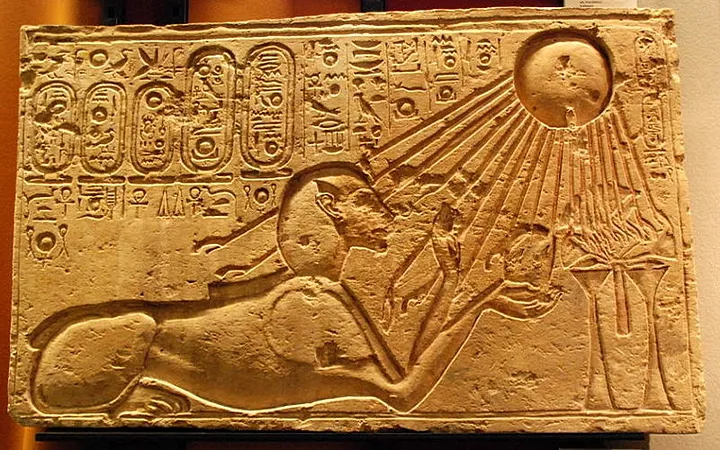
  • Pharaoh Akhenaten (18th dynasty) apparently had a hydrocephalic skull, possibly symbolic artificial deformation; his daughters’ heads returned to normal depiction posthumously.akhenatenPharaoh Akhenaten
  • Hippocrates (466-377 BC) described hydrocephalus as a consequence of epilepsy:
    • "When epilepsy has become chronic it is no more curable. The brain is corroded by pituita and melts, forming water that surrounds the remaining brain outside."
    • Noted clinical symptoms: headache, diplopia, impaired visual function, vomiting.
    • Recommended treatments included vegetables, sneeze-inducing inhalants, and ultimately trephination.
  • Ancient physicians distinguished four types of hydrocephalus, as recounted by Paré.
  • Hydrocephalic children were viewed as monstrous and often abandoned or starved.
  • Celsus (25 BC - AD 37), in "De medicina", described extracranial hydrocephalus:
    • Skin elevated by fluid forming a soft, tumour-like swelling responsive to finger pressure.
    • Greek term "hydrokephalos" applied to extracranial fluid accumulation; intracranial treatment considered too risky.
  • Paulus of Aegina (7th century), summarized hydrocephalus as an accumulation of watery fluid in his "Compendii (medici) libri septem".
  • Galen (AD 131-201) described four types of hydrocephalus:
    • Between brain and meninges
    • Between meninges and bone
    • Between bone and pericranium
    • Between pericranium and skin
    • Declared only extracranial type (between skin and pericranium) as curable via incision; intracranial type incurable.
  • Ambroise Paré (1500-1590), in "De l'eau qui vient à la teste des enfans, appellee Hydrocéphale", identified four fluid accumulation sites:
    • Between skin and pericranium
    • Between pericranium and skull
    • Between skull and dura mater
    • Within brain ventricles and substance

Fig 1

ambroise pare hydrocephalus 1

Post-Christian Era

Roman-Byzantine Medicine

  • Among prominent Byzantine physicians (such as Aëtius of Amida, Alexander of Tralles, and Paulus of Aegina), Paulus (7th century AD) summarized earlier medical knowledge in his work "Compendii medici libri septem", referencing Hippocrates, Galen, Celsus, Antyllus, and Aëtius.
  • Paulus described hydrocephalus as a childhood condition characterized by an accumulation of watery fluid. He distinguished between extracranial forms and accumulations situated between the skull and meninges, with fluid potentially escaping through widened cranial sutures. Clinical signs included frontal enlargement of the head, fixed eyes, and tears. He advised against surgical intervention due to high risk.
  • Oribasius (4th century AD), another Byzantine physician, identified three types of hydrocephalus:
    • Accumulation below the scalp (surgically treatable).
    • Accumulation below the pericranium (surgically treatable).
    • Accumulation between the skull and meninges (considered incurable).

Islamic Medicine

  • Between AD 900–1100, Islamic physicians such as Rhazes, Avicenna, Ali Abbas, and Albucasis extensively characterized hydrocephalus.
  • Rhazes recommended compression therapy, while Avicenna suggested cauterization of the cranial sutures.
  • Albucasis (AD 1013), renowned for his surgical approaches, thoroughly detailed hydrocephalus in his Latin-transcribed work "Chirurgicorum omnium Primarii lib. tres". He noted that hydrocephalus typically occurred in infancy, due either to birth trauma or unknown causes. He described a case at autopsy identifying a congenital obstruction of cerebrospinal fluid drainage, advising against surgical intervention unless fluid accumulation occurred extracranially or just beneath the skull.
  • Albucasis recommended precise surgical techniques:
    • Extracranial cases: incision with a scalpel.
    • Subcranial (external) cases: three-armed incision followed by careful covering and application of ointment.

albucasis scalpelScalpel used by Albucasis 

  • Albucasis cautioned strongly against accidental arterial damage, emphasizing the risk of fatal hemorrhage during fluid evacuation procedures.
  • Ali Fárábí (AD 870–950), the prominent Aristotelian scholar, noted the frequent occurrence of hydrocephalus in sheep, indicating a broad awareness of this condition in animals as well (cited in Paulus Ægineta, AD 1846).

European Renaissance

  • The influence of Christianity, especially after the Church's ascendancy, significantly impacted the interpretation of congenital abnormalities, including hydrocephalus. Augustine (AD 354–430), in "De Civitate Dei," described various human deformities ("cripples with heads of dogs," cyclopes, monsters) but emphasized that every being possessed a mortal soul, arguing against contemporary beliefs that killing such individuals was justified. Augustine's views and Roman law influenced subsequent Christian attitudes toward congenital malformations.
  • In AD 1140, Gratian's Canonical Laws stipulated that children born with severe deformities (considered omens of war, misfortune, or plague) should not be killed, marking a significant ethical shift from previous practices.
  • With the advent of printing in the 15th century, Renaissance literature increasingly documented teratological conditions. Notable works included "De occultis naturae miraculis libri duo" (1559) by Levinus Lemnius (1505–1568, Netherlands) and "De Monstrorum Natura" (1634) by Fortunio Licetus (1577–1657, Italy), which described various congenital anomalies.
  • Hieronymus Mercurialis (1530–1606) from Padua, in his "De morbis puerorum" (1583), detailed internal hydrocephalus associated with apoplexy and epilepsy, attributing its cause to uterine overheating and recommending Galenical treatments.
  • Leonardo da Vinci accurately depicted the structure of the ventricular system in 1505, using wax casts from an ox brain.
  • Andreas Vesalius (1514–1564), in his seminal work "Fabrica Libri Septem" (1543), described a distinctive skull deformation similar to Hippocrates' earlier descriptions. Vesalius observed a case in Venice—a mentally impaired boy with pronounced skull deformation—and noted similar anomalies in other cases from Bologna and Genoa. He asserted that unusual skull shapes were occasionally found even in intellectually accomplished individuals ("egregie prudentibus").
  • Vesalius also extensively documented a detailed case of hydrocephalus in a 2-year-old girl from Augsburg (1551). Within seven months, her head expanded dramatically. Remarkably, despite significant ventricular dilation ("three Augsburg wine measures"), the child retained sensory and motor functions until her final days. Vesalius expressed astonishment at the absence of severe neurological impairment despite substantial fluid accumulation.
  • In the 16th century, the trephining procedure for external hydrocephalus ("Observations et Histoires Chirurgiques," 1679) became controversial. Some physicians, such as De Villeneuve, recommended trephining for vertigo; others like Roland (died 1562), Guillaume Rondelet (1507–1566), and Petrus Forestus (1522–1597) advocated it for mental illness, while Jacques Hollier and Marcellus Donatus used it for epilepsy. Prominent medical scholars, including Giovanni Ingrassia (1510–1580, Palermo) and Guilio Aranzi (1530–1589, Bologna), considered such surgical interventions too risky.
  • Jean Fernel (1499–1558), a renowned French court physician, advanced understanding of brain physiology. In Portugal, Amatus Lusitanus (1511–1561), a prominent converted Jewish physician, famously performed surgery on a hydrocéphalie mole, attracting significant attention.
  • Despite advancements in anatomical knowledge, internal hydrocephalus remained widely viewed as surgically untreatable during this period, with management limited to head wrapping and Galenical remedies.

The 17th Century

  • The Danish anatomist Niels Stensen (1638–86) did not frequently use references, except in his "Chaos manuscript". It is therefore advantageous to review the literature prior to his letter to Duke Fernando II of Tuscany describing the autopsy of a hydrocephalic calf in 1669 in Innsbruck.

ventricular system hydrocephalus calf stensenThe ventricular system of a hydrocéphalic calf (Stensen 1669)

  • In 1600, a son of Madame Clarice Caraffe was described. By the age of 5, he had developed a skull tumor, which four years later reached substantial size, spanning two thumbs and five fingers across (Severino, 1668; "Observations et Histoires Chirurgiques", 1679). The tumor was thought to result from blocked cerebrospinal fluid flow, causing bulging of skull sutures and prominent swelling in the right temporal region. Clinical signs included deep-set eyes with limited downward movement, head ulceration, tooth loss, cognitive impairment, and eventual death.

severino 5yr boy hydrocephalus

  • Marco Aurelio Severino (1580–1656), a contemporary and close associate of the renowned anatomist Thomas Bartholin (1616–70) of Copenhagen, documented descriptions by Albucasis, Rhazes, and Hildanus (1560–1634) as early as 1606. Severino noted hydrocephalus commonly occurred in robust parents' offspring, possibly originating around the seventh embryonic month due to uterine overheating. Despite cranial enlargement and clear intracranial fluid accumulation, posterior brain regions and cerebellum frequently remained unaffected. Severino offered no treatment recommendations (Severino 1629, lib. IV, cap. 9 & 12).
  • Many 17th-century texts continued using older classifications distinguishing extra- and intracranial forms of hydrocephalus. Prominent authors included Germans Fabricius Hildanus (1560–1634), C.V. Schneider (1614–1680), G. Moebius (1611–1667), and Daniel Sennert (1572–1637). Other notable contributors were Nicolaas Tulp (1593–1674) of the Netherlands (depicted famously by Rembrandt), Zacutus, Jan van Home (Stensen's teacher), Pierre Borel (1620–1671) of France, De Aquapenden of Italy, and Thomas Willis (1621–1675) and Francis Glisson (1597–1677) from England.
  • Willis (1664) proposed that brain excretions flowed into ventricles, hypothesizing the pineal gland or choroid plexus as fluid-producing structures.
  • The French patho-anatomist Theophile Bonet (1620–89) referenced Danish work on hydrocephalus in "Medicina Septentrionalis" (1684), citing Thomas Bartholin (1616–1680), physician Frederik Hammerich (1628–83) from Elsinore, Copenhagen barber-surgeon Nicolai Boye (1615–1690), and chemist Oluf Borck (1626–1690).

Fig 1

theophili boneti

  • Bartholin notably mentored Stensen, describing in 1654 a 5-year-old hydrocephalic boy whose head matched the circumference of three adults. Despite severe deformities limiting speech and mobility, his eleven siblings were unaffected.

bartholin hydrocephalus

  • During Stensen's tenure in Leyden alongside his mentors Franciscus de la Boë Sylvius (1614–1672)—who first described the cerebral aqueduct in 1641—and Jan van Home (1598–1649), he pursued rigorous anatomical and pathological brain studies, including extensive investigations on the parotid gland and heart.
  • Sylvius did not publish his hydrocephalus findings, but Leyden surgeon Paul Barbette’s 1663 work on hydrocephalus and the cerebral aqueduct notably reignited Stensen's research interest.

barbette hydrocephalus

  • In 1665, Stensen delivered a landmark Paris lecture titled "The Anatomy of the Brain," subsequently performing autopsy on a turtle exhibiting hydrocephalus.
  • Stensen's pivotal 1669 dissection of a hydrocephalic calf concluded that hydrocephalus resulted from a tumor obstructing cerebrospinal fluid drainage, rather than maternal illness. He identified remnants near the optic chiasm, suggesting a craniopharyngioma. Detailed examination revealed significantly enlarged lateral ventricles holding approximately four pounds of fluid, exerting pressure at points of least resistance.
  • Stensen emphasized remarkable preservation of motor and sensory functions despite severe cerebral compression, offering one of the earliest pathophysiological explanations of hydrocephalus. Detailed anatomical illustrations accompanied his findings (Stensen, 1669).

Late 17th, 18th and 19th Centuries

  • In the late 17th century, patho-anatomical research flourished, bringing scientific attention to hydrocephalus and skull deformities. The Galenic belief of a vaporous brain substance persisted into this period.
  • Thomas Willis (1664) hypothesized that cerebrospinal fluid (CSF) originated from the pineal gland or choroid plexus. Albrecht von Haller (1708–1777), however, argued the ventricles contained a vaporous substance during life, condensing to fluid post-mortem.
  • Théophile Bonet (1620–1689) of Geneva systematically documented medical cases in his influential review "Sepulchretum" (1679). This collection included approximately 3000 cases from 470 authors. Bonet specifically described hydrocephalus ("Hydrocephalus sive hydrope capitis") in 16 detailed observations, referencing works by Vesalius, Wepfer, Schneider, and Hildanus.
  • In 1684, Bonet published "Medicina Septentrionalis", which featured a detailed description of hydrocephalus, notably illustrating Stensen's famous "Vitulus hydrocephalus" (hydrocephalic calf).
  • The Italian patho-anatomist Giovanni Morgagni (1682–1771) described hydrocephalic brains as reduced to a thin membrane-like structure. Morgagni warned that tapping such brains could misleadingly suggest near-perforation of skull and dura, and also described hydrocephalus in adults.
  • In 1768, Robert Whytt (1714–1766) of Edinburgh published an outstanding work titled "Observations on the Dropsy in the Brain", characterizing tuberculous meningitis and communicating hydrocephalus. Whytt divided hydrocephalus into three stages based on clinical progression and documented clear fluid accumulation immediately below the corpus callosum in autopsied cases.

whytt dropsy brain

  • Dominico Cotugno (1736–1822) confirmed the existence of spinal CSF in 1764 and suggested that it could be accessed via percutaneous tap. He disproved Galen's vapor theory, establishing definitively that the ventricles contained fluid rather than vapor.
  • Nearly a century later, another Danish researcher, N.D. Riegels (1755–1802), described a hydrocephalic calf in a meticulously hand-written Latin manuscript titled "Dissectio vitulae sine cerebro" (1793). Riegels referred extensively to earlier research by Tulp, du Hamel, Stensen, Morgagni, and Littré, though notably omitted Robert Whytt's influential findings regarding spinal cord integrity from 1750. Riegels extensively quoted prominent physicians such as Boerhaave, Morgagni, von Haller, Petit, and Le Dran in his reflections on internal hydrocephalus.

calf dissectionThe dissection of the head of a calf (Riegels, 1793)

  • Joseph Lieutaud, a French royal court physician, continued to emphasize traditional humoral theory in his synopsis "Universae Praxeos Medicae" (1765). William Buchan (1729–1803) in "Domestic Medicine" (1786) also maintained the distinction between external (treatable) and internal (untreatable) hydrocephalus, reflecting the persistent influence of humoral concepts.
  • In 1789, Michael Underwood (1737–1820) notably published "A Treatise on the Diseases of Children", clearly distinguishing external hydrocephalus from internal ventricular hydrocephalus. He observed that internal hydrocephalus could permit longer survival, whereas fluid accumulation external to the brain surface rapidly proved fatal. Underwood recommended therapeutic application of vesicants over the superior sagittal sinus.

The 19th Century

  • In 1828, Danish physician Christian de Meza (1756–1844) reported a "case of hydrocephalus combined with rupture of the abdomen."
  • In 1852, Harald Selmer (1814–1879) described a notable case of congenital hydrocephalus in a 15-year-old boy.
  • Hans Sommerfeldt (1804–1867) defended his thesis "De Hydrocephalo Acuto" in 1836, providing an extensive and valuable account of acute hydrocephalus between 1770 and 1830. Sommerfeldt referenced Robert Whytt's work, listed common therapeutic drugs (including mercury preparations, laxatives, diuretics, and digitalis), and classified the disease under several terminologies such as "Hydrocephalitis," "Hydrops cerebri," "Hydrops ventriculorum cerebri," "Encephalitis enfantum," and "Apoplexia hydrocephalia." He emphasized the uniformly poor prognosis ("prognosis pessima") associated with these conditions.
  • Johannes Starck (1799–1873), a Danish barber-surgeon, defended his thesis "De Hydrocephali Paracentesi" in Rostock in 1841, exploring infantile hydrocephalus and potential surgical interventions. Starck distinguished various hydrocephalic forms, particularly identifying "hydrocephalus chronicum," corresponding closely to today's definition of chronic hydrocephalus.
  • Starck conducted a meticulous comparison between proponents and opponents of surgical intervention for chronic hydrocephalus (hydrocephalus chronicum sive hydrops ventriculorum):
    • Supporters of surgery included René Descartes (1689), Liones (1716), Desauvages (1768), and Alexander Monro III (1821). In cases of delayed childbirth, surgery was advocated by Wildanus (1721), Erasmus Darwin (1797), and Heinrich Callisen (1817).
    • Opponents of surgery included historical figures such as Aëtius, Ambroise Paré, Herman Boerhaave (1738), Martini (1769), Giovanni Morgagni (1761), and the prominent hydrocephalus researcher Leopold R. Gölis (1765–1827).
  • Starck’s thesis notably avoided proposing specific hypotheses or recommendations concerning surgical intervention. Though he published no further research specifically on hydrocephalus, Starck became well-known in Denmark for pioneering advancements in tenotomies and plastic surgery.
  • In 1848, John Cheyne (1777–1836) and Charles West (1816–1898) classified hydrocephalus into acute and chronic forms, documented brain malformations, and identified acquired hydrocephalus secondary to tumors, hemorrhage, and meningitis.
  • François Magendie (1783–1855), in 1825, reported the midline opening of the fourth ventricle and described cerebrospinal fluid (CSF) movement. Herbert von Luschka (1820–1875) subsequently identified the lateral openings (foramina of Luschka) in the fourth ventricle in 1855. These anatomical insights led Swedish anatomists E.A.H. Key & G. Retzius in 1875 to comprehensively illustrate CSF pathways and absorption mechanisms in their influential anatomical atlas, thoroughly detailing the subarachnoid spaces.
  • In 1837, J. Coster, in his "Handbook of Practical Medicine," criticized conventional mercurial treatments of hydrocephalus as ineffective, advocating instead for prompt general bleeding. Coster cautioned strongly against trephining, noting all known cases ended fatally.
  • Pensa (1840) extended Underwood’s work, differentiating acute and chronic hydrocephalus. Acute hydrocephalus was linked to inflammatory processes, supported by authors like Rusch, Temple, Fercival, and Paterson (di Rocco, 1987). Despite aggressive anti-inflammatory therapy, outcomes remained uniformly fatal. Chronic hydrocephalus offered slightly better prognostic hopes, leading to a tentative classification into three subtypes.
  • Joseph François Malgaigne (1806–1865), in his 1834 "Manuel de médecine opératoire," described ventricular punctures as questionable procedures. However, influenced by John Conquest (1789–1866)—a London obstetrician who reported success in 10 out of 19 hydrocephalus cases treated by puncture (1838)—Malgaigne later reconsidered his stance.
  • James R. Bennett reviewed medical approaches to hydrocephalus in 1843. Interest in hydrocephalus treatment surged, driven by physiopathological insights from Magendie and Key & Retzius on CSF dynamics.
  • Magendie notably described thrombosis and occlusion of the vein of Galen as a pathogenic mechanism for hydrocephalus, attributing increased CSF volume to reduced absorption resulting from venous obstruction.
  • Key & Retzius’s work significantly advanced the understanding of CSF absorption into the venous sinuses. This discovery profoundly impacted hydrocephalus management by providing a rational framework for ventricular dilatation across various hydrocephalic presentations.
  • In 1879, John Hilton (1804–1878), author of "Rest and Pain" (1863), introduced the concept of mechanical obstruction as the cause of ventricular dilation, prompting a wave of surgical interventions.
  • Extensive surgical reviews on hydrocephalus by Davidoff (1929), Ring-Mrozik & Angerpointner (1986), and Di Rocco (1987) described varied puncture techniques performed throughout the 19th century by surgeons such as Conquest, Graefe, Russell, Malgaigne, Langebask & Hahn, and Bouchout & Chipault. Di Rocco detailed diverse puncture sites (fontanelle, orbital roof, nostrils), depths, and volumes drained, ranging from 50–250 ml per procedure, up to 1500 ml from two punctures (Malgaigne, 1849).
  • Numerous extrathecal shunts were attempted from ventricles, cisterna magna, or lumbar subarachnoid space. Heinrich Quincke (1842–1922) notably advocated repeated lumbar punctures in 1891 as less hazardous for patients, pioneering this safer palliative approach.

The 20th Century

Europe

  • In 1909, Professor Thorkild Rovsing (1862–1927), at the University of Copenhagen, published a significant report on the surgical treatment of hydrocephalus. Around the same time, Professor E.A. Tscherning (1851–1919), at The Municipal Hospital of Copenhagen, described bilateral ventricular drainage procedures.
  • In 1911, Rovsing further detailed a technique involving permanent communication between the lateral ventricles and the subarachnoid space using guttapercha-paper drainage. Rovsing reported good clinical outcomes with significant symptom improvement.
  • Apart from Albucasis, operative interventions for hydrocephalus were generally discouraged for approximately one thousand years. Conventional treatments typically included head wrapping, medications, and bloodletting (phlebotomy).
  • Around the beginning of the 20th century, however, several European surgeons started advocating operative treatments. Ballance (1908) successfully described techniques involving intradural drainage from the ventricles into the subdural space using small, angled tubes made from gold, iridium, or platinum.
  • In 1908, W. Kausch pioneered a method for shunting cerebrospinal fluid (CSF) from the lateral ventricles into the abdominal cavity. Ballance also described carotid artery ligation to decrease CSF secretion in cases of congenital hydrocephalus.

USA

  • Lewis Weed (1886–1956), Walter Dandy (1886–1946), and Kenneth Blackfan (1883–1941) became pioneering American neurosurgeons who initiated critical research into the physiology and circulation of CSF in 1914, particularly investigating hydrocephalus.
  • They developed experimental hydrocephalus models in dogs by obstructing the aqueduct of Sylvius, demonstrating ventricular dilation proximal to the obstruction.
  • Dandy and Blackfan demonstrated that ventricles without choroid plexuses did not dilate when obstructed at the foramen of Monro, whereas ventricles containing a choroid plexus expanded significantly. Their research reinforced the longstanding belief that choroid plexuses primarily produced CSF.
  • Walter Dandy's introduction of ventriculography using air as a contrast medium in 1918 significantly advanced hydrocephalus diagnostics. This breakthrough spurred surgical innovations, notably plexectomy procedures (either direct removal or cauterization as advocated by Harvey Cushing in 1926), primarily effective for communicating hydrocephalus.

The Era 1930–1960

  • In 1929, Leo Max Davidoff (1898–) provided an influential historical overview of hydrocephalus, highlighting improved understanding of cerebrospinal fluid (CSF) dynamics and pathology.
  • In 1938, A. Thorkildsen developed the ventriculo-cisternal shunt, and later, Stookey & Scharff introduced third ventriculostomy as alternative surgical treatments.
  • Further research during this period demonstrated additional sources of CSF production beyond the choroid plexus. Approximately 40% of CSF was found to originate directly from brain tissue itself (Davson 1967, Bering 1966, Pollay 1967, Welch 1966, Milhorat 1972).
  • With advancements in neurosurgery across Europe and the United States, innovative methods emerged for CSF diversion, including shunting to diverse anatomical locations such as the heart, jugular vein, ureter, pleural space, Fallopian tube, mastoid air cells, sagittal sinus, intestinal tract, gall bladder, spinal epidural space, thoracic duct, and even the parotid gland (Scharff 1963, Milhorat 1972).
  • A significant advancement occurred in 1952 when Nulsen & Spitz, closely followed by Holter, developed the one-way valve shunt system, enabling effective drainage of CSF directly into the jugular vein. This innovation led to the development of numerous specialized shunt devices and various operative techniques.
  • In contemporary practice, the use of third ventriculostomy and ventriculo-cisternostomy has become less common, while ventriculo-atrial, ventriculo-peritoneal, or lumbo-peritoneal shunts remain the preferred surgical methods (Guertin, 1987).

The Era 1960–1990

  • The classification of hydrocephalus introduced by Dandy & Blackfan (1914) remains relevant, though significantly enriched by extensive new experimental and clinical insights (Wolstenholme and O'Connor, 1958).
  • During this period, hydrocephalus classifications expanded considerably, including:
    • Age-based: congenital, juvenile, and adult hydrocephalus.
    • Aetiological: congenital, acquired, and unknown causes.
    • Clinical status: active or arrested hydrocephalus.
    • Patho-anatomical or investigative findings: non-communicating (obstructive) and communicating (acquired adhesions) hydrocephalus.
    • Pathophysiological (ICP monitoring): hydrocephalus with increased or normal intracranial pressure (ICP).
    • CSF dynamic findings: increased resistance to CSF outflow versus normal resistance.
    • Computed tomography (CT) imaging: patterns based on ventricular size, periventricular lucency, and size of cerebral sulci.
  • Previously, CSF dynamics were assessed using isotope cisternography through lumbar or ventricular injections; however, these techniques yielded limited value for evaluating post-shunt outcomes (Borgesen, 1984).
  • The last three decades notably saw significant advancements in:
    • Development and refinement of various shunt systems.
    • Measurement techniques for intracranial pressure (ICP) and resistance to CSF outflow (Rout).
    • Enhanced understanding of hydrocephalus pathophysiology, particularly normal pressure hydrocephalus.
  • Investigation strategies for newly diagnosed hydrocephalus in both children and adults have been structured into systematic decision trees.
  • Research into the "third circulation" (CSF circulation) in recent decades emphasized:
    • Secretion and biochemical composition of CSF.
    • Classification and management of various hydrocephalus types.
    • CSF absorption and accurate measurement of resistance to CSF outflow.
    • Pathophysiological roles of CSF-transmitters and peptides.
  • Comprehensive reviews and reference volumes extensively covered these significant advancements (e.g., Borgesen, Milhorat, and others).

Further reading

Gjerris, F., & Snorrason, E. (1992). The history of hydrocephalus. Journal of the History of the Neurosciences1(4), 285–312. https://doi.org/10.1080/09647049209525541


Cover Image: Relic found among the ruins of Amarna. It depicts Pharaoh Akhenaten as a Sphinx — a guardian spirit — touched by the sacred rays of the god Aten, who is depicted as a sun disk with rays that end in hands (image source: Wikimedia Commons)